Cardiac imaging has become a cornerstone of modern cardiology, empowering physicians to see the heart in ways that were once unimaginable. Through continuous innovation and the integration of advanced technology, cardiac imaging now plays a vital role in diagnosing, monitoring, and guiding treatment for a wide range of heart conditions. These advancements allow for earlier detection, more accurate assessments, and personalized care tailored to each patient’s unique heart profile.
Revolutionary Echocardiography Techniques
Echocardiography remains one of the most versatile and widely used imaging tools in cardiology. Modern advancements such as 3D echocardiography and stress echocardiography have elevated the precision and clarity of cardiac evaluation. These technologies provide real-time visualization of the heart’s chambers, valves, and blood flow, helping physicians detect issues such as valve dysfunction, muscle weakness, or fluid buildup. The dynamic images offer a deeper understanding of cardiac function under stress, guiding decisions on medical therapy or intervention.
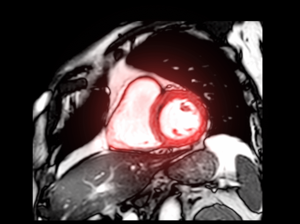
Cardiac MRI delivers high-resolution, detailed images of the heart’s structure and tissue composition. It enables the detection of myocardial scarring, inflammation, congenital abnormalities, and other subtle changes often missed by conventional imaging. This makes it an invaluable tool for personalized treatment planning, especially in patients with complex or chronic heart conditions. By providing a clear view of how the heart muscle is functioning, MRI helps tailor therapies for better long-term outcomes.
CT coronary angiography has transformed the way physicians visualize coronary arteries. This non-invasive imaging technique can detect plaque buildup, arterial narrowing, and early signs of coronary artery disease with remarkable accuracy. For many patients, it eliminates the need for invasive catheter-based procedures, reducing both recovery time and potential complications. The integration of AI-based image analysis further enhances detection speed and diagnostic precision.
The clearer we see the heart, the closer we come to healing it.
Nuclear imaging methods such as PET (Positron Emission Tomography) and SPECT (Single Photon Emission Computed Tomography) allow doctors to assess blood flow, perfusion, and metabolic activity in heart tissues. These scans help identify areas at risk of ischemia or tissue damage, especially after heart attacks. They also assist in monitoring the effectiveness of treatments and predicting recovery outcomes, ensuring that therapy is both targeted and efficient.
Guiding Interventions and Enhancing Outcomes
Modern cardiac imaging does more than just diagnose—it actively guides interventional and surgical procedures. By providing precise anatomical mapping, imaging helps cardiologists and surgeons plan every step of a procedure with confidence. Whether it’s stent placement, valve repair, or minimally invasive surgery, imaging technologies ensure accuracy, minimize risk, and enhance patient recovery.

Beyond its clinical benefits, cardiac imaging plays a crucial role in patient education and engagement. Seeing their own heart images helps patients understand their condition better, recognize the importance of follow-up care, and stay committed to lifestyle and medication plans. Transparent communication between patients and physicians enhances trust and encourages proactive participation in health management.